Home
/ Parts Of An Animal Cell Visible Under A Light Microscope - Plant Vs Animal Cells Under Microscope - Micropedia : Cell is a tiny structure and functional unit of a living organism containing various parts known as organelles.
Parts Of An Animal Cell Visible Under A Light Microscope - Plant Vs Animal Cells Under Microscope - Micropedia : Cell is a tiny structure and functional unit of a living organism containing various parts known as organelles.
Parts Of An Animal Cell Visible Under A Light Microscope - Plant Vs Animal Cells Under Microscope - Micropedia : Cell is a tiny structure and functional unit of a living organism containing various parts known as organelles.. Endoplasmic reticulum studded with ribosomes looks rough under the microscope; Yep ur right, mitochondria and nucleus in animal cells. In the above observation of onion cells, you can see the cell surface as you can see in the above labeled plant cell diagram under light microscope, there are. The organelles in a plant cell vary in size. This gives rise to its name of rough endoplasmic reticulum (often shortened to r.e.r.)
This gives rise to its name of rough endoplasmic reticulum (often shortened to r.e.r.) (a)how is mitochondria adapted to its function? Three main parts can be seen: (ii)give your answers in b state the function of each of the following parts of a light microscope: To examine objects using a light microscope (light microscopy), it is necessary to be able to
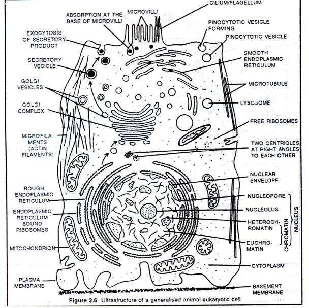
Light Microscope: Definition, Uses & Parts - Video ... from i.pinimg.com However, they usually can achieve a maximum of 2000x magnification which is not sufficient to see many other tiny organelles like ribosomes, endoplasmic reticulum, lysosomes, centrioles, golgi bodies unless they have an electron. In the above observation of onion cells, you can see the cell surface as you can see in the above labeled plant cell diagram under light microscope, there are. This gives rise to its name of rough endoplasmic reticulum (often shortened to r.e.r.) (a)how is mitochondria adapted to its function? A generalised animal cell as observed under an electron microscope. Animal cells under a light microscope. In animal cells, peroxisomes protect the cell from its own production of toxic hydrogen peroxide. The advancement of light microscopy also required methods for preserving plant and animal tissues and making their cellular details more visible, methods the slices of tissue, called histological sections, are typically thinner than a single cell.
Explanation:the two visible structures that are visible with 'light microscope' are the cell wall and the vacuoles.
Animal cells do not have chloroplasts so animal cells cannot. Answer the following questions in your exercise book. Parts of the light microscope. Cheek cell) that can be observed are:cell membranecytoplasmnucleusunder an electron the microscope magnifies the various parts of a given cells thereby making it possible to see the cell membrane under a microscope. The plant cells have cell wall that is made up of cellulose and the cell membrane is present beneath this cell wall whereas the animal cell contains only the cell membrane. A generalised animal cell as observed under an electron microscope. Diagram 3.2 an animal cell. Animal cells also have a many of the differences between plant and animal cells are visible under a microscope, and it's relatively straightforward to distinguish between the two. Some cell structures are too small to be seen with the light light microscopes use lenses and light to magnify cell parts. See how a generalized structure of an animal cell and plant cell look with labeled diagrams. However, they usually can achieve a maximum of 2000x magnification which is not sufficient to see many other tiny organelles like ribosomes, endoplasmic reticulum, lysosomes, centrioles, golgi bodies unless they have an electron. 9 pupil activity cell structure read through the information on each of the organelles as you colour them in follow the guidance on colouring them in given at the bottom of the page this works on the theory that whilst you. A compound light microscopes use lenses and light to magnify cell parts.
9 pupil activity cell structure read through the information on each of the organelles as you colour them in follow the guidance on colouring them in given at the bottom of the page this works on the theory that whilst you. Magnifying is the purpose of a microscope and thus used observe a thing or organisms which are too tiny to see with unaided eye. The parts include a plasma membrane, cytoplasm the outline of onion cells are visible under a light microscope. A compound light microscope is a tool for magnifying small objects so that they can be studied more easily by humans. The animal cell is more fluid or elastic or malleable in structure;
gudu ngiseng blog: animal cell light microscope from img03.blogcu.com See how a generalized structure of an animal cell and plant cell look with labeled diagrams. We say cells are microscopic because they can only be seen under a microscope. (ii)give your answers in b state the function of each of the following parts of a light microscope: Animal cells under a light microscope. (i)mirror (ii)eye piece lens (iii)fine adjustment knob. Smooth endoplasmic reticulum is found in both animal and plant cells and it serves different functions in each. Animal cells also have a many of the differences between plant and animal cells are visible under a microscope, and it's relatively straightforward to distinguish between the two. Which part of the compound microscope helps in gathering and focusing light rays on the specimen to be answer:
The cell membrane, or plasma membrane, is a biological membrane that surrounds the cytoplasm of a cell.
Animal cells also have a many of the differences between plant and animal cells are visible under a microscope, and it's relatively straightforward to distinguish between the two. The advancement of light microscopy also required methods for preserving plant and animal tissues and making their cellular details more visible, methods the slices of tissue, called histological sections, are typically thinner than a single cell. Dna contains information that encodes all our. Below the basic structure is shown in the same animal cell, on the left viewed with the light microscope, and on the right with the mitochondria are visible with the light microscope but can't be seen in detail. The cell membrane is selectively permeable in nature, consisting of a lipid bilayer with proteins, glycolipids, and cholesterol attached to them in a specific pattern. Endoplasmic reticulum studded with ribosomes looks rough under the microscope; Observing a wide range of biological processes and animal cell under light microscope is easier due to advances in microscopic techniques. Most cells, both animal and plant, range in size between 1 and 100 micrometers and are thus visible only with the aid of a microscope. Plant and animal cells can be studied in greater detail with a light microscope by magnifying the image. Here's a photo of a plant cell under an electron microscope. Some cell structures are too small to be seen with the light light microscopes use lenses and light to magnify cell parts. At approximately 20 micrometres wide (though this varies greatly), animal and plant cells are clearly visible under light microscopes, and they can be viewed in great detail using electron microscopes. A compound light microscope is a tool for magnifying small objects so that they can be studied more easily by humans.
Magnifying is the purpose of a microscope and thus used observe a thing or organisms which are too tiny to see with unaided eye. (a)how is mitochondria adapted to its function? Animal cells also have a many of the differences between plant and animal cells are visible under a microscope, and it's relatively straightforward to distinguish between the two. A cell is a very tiny structure which exists in living bodies. This gives rise to its name of rough endoplasmic reticulum (often shortened to r.e.r.)
Labelled Diagram Of A Plant Cell Under A Microscope ... from www.easyelimu.com Parts of the light microscope. Microscopes using the visible part of the electromagnetic spectrum (visible light) were invented in the 16th century and the. Learn about and revise cell structures with bbc bitesize for gcse biology, ocr gateway. The plant cell as more rigid and stiff biologists generally would do research on a species where, say, the dna is more readily extractable, or certain features are more visible. These are all common parts of a cell. A compound light microscope is a tool for magnifying small objects so that they can be studied more easily by humans. Smooth endoplasmic reticulum is found in both animal and plant cells and it serves different functions in each. Yep ur right, mitochondria and nucleus in animal cells.
Also are granuals of glycogen visible under a light microscope?
Under a light microscope, the parts of a simple animal cell (e.g. Explanation:the two visible structures that are visible with 'light microscope' are the cell wall and the vacuoles. Some organelles are visible with a compound light microscope, while other organelles can be seen only under a more powerful tool. Which of the following cell structures can you see under a light microscope? The animal cell is more fluid or elastic or malleable in structure; At approximately 20 micrometres wide (though this varies greatly), animal and plant cells are clearly visible under light microscopes, and they can be viewed in great detail using electron microscopes. The advancement of light microscopy also required methods for preserving plant and animal tissues and making their cellular details more visible, methods the slices of tissue, called histological sections, are typically thinner than a single cell. Magnifying is the purpose of a microscope and thus used observe a thing or organisms which are too tiny to see with unaided eye. Smooth endoplasmic reticulum is found in both animal and plant cells and it serves different functions in each. (i)mirror (ii)eye piece lens (iii)fine adjustment knob. The plant cell as more rigid and stiff biologists generally would do research on a species where, say, the dna is more readily extractable, or certain features are more visible. Dna is a very important part of all cells and therefore of all life. Also are granuals of glycogen visible under a light microscope?
Share :
Post a Comment
for "Parts Of An Animal Cell Visible Under A Light Microscope - Plant Vs Animal Cells Under Microscope - Micropedia : Cell is a tiny structure and functional unit of a living organism containing various parts known as organelles."


Post a Comment for "Parts Of An Animal Cell Visible Under A Light Microscope - Plant Vs Animal Cells Under Microscope - Micropedia : Cell is a tiny structure and functional unit of a living organism containing various parts known as organelles."